
Ultrasound Imaging Needle
January 30, 2018 | Terry Sharrer
Optical Ultrasound of the Heart
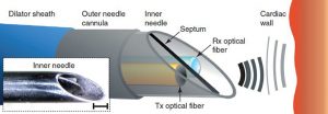
For the first time, it has become possible to make live images from inside the heart during surgery, by means of an optical ultrasound “needle.” “The needle system can emit ultrasound thanks to the development of a novel composite material consisting of a mesh of carbon nanotubes encased in silicone and located on the tip of the optical fiber. The pulsed light from the fiber is absorbed by the carbon nanotubes and produces an ultrasound wave due to the photoacoustic effect. A second innovation underpins the detection of the reflected ultrasound waves at such a small scale, which is the invention of highly sensitive optical fibers incorporating polymer optical microresonators.” MORE WITH VIDEO
Image Credit: University College London and MedGadget.com
