
Improved Imaging of Arterial Plaques
Improved imaging of arterial plaques
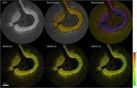
“Here we [at the University of California-Davis, and Massachusetts General Hospital] report the first implementation of an intravascular catheter system combining fluorescence lifetime imaging (FLIm) with polarization-sensitive optical coherence tomography (PSOCT). This system provides multi-scale assessment of plaque structure and composition via high spatial resolution morphology from OCT, polarimetry-derived tissue microstructure, and biochemical composition from FLIm, without requiring any molecular contrast agent. This result was achieved with a low profile (2.7 Fr) double-clad fiber (DCF) catheter and high speed (100 fps B-scan rate, 40 mm/s pullback speed) console. . . . The unique ability to collect complementary information from tissue (e.g., morphology, extracellular matrix composition, inflammation) with a device suitable for percutaneous coronary intervention offers new opportunities for cardiovascular research and clinical diagnosis.” MORE
Image Credit: EurekaAlert
