
Imaging the Brain’s Oxygen
Real-time whole-brain imaging of hemodynamics and oxygenation at micro-vessel resolution with UFF-PAM
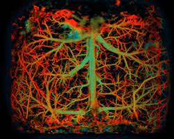
“Here, we [at Duke University] present a novel ultrafast functional photoacoustic microscopy (UFF-PAM) to image the whole-brain hemodynamics and oxygenation. UFF-PAM takes advantage of several key engineering innovations, including stimulated Raman scattering (SRS) based dual-wavelength laser excitation, water-immersible 12-facet-polygon scanner, high-sensitivity ultrasound transducer, and deep-learning-based image upsampling. . . . Using the UFF-PAM system, we have demonstrated proof-of-concept studies on the mouse brains in response to systemic hypoxia, sodium nitroprusside, and stroke. We observed the mouse brain’s fast morphological and functional changes over the entire cortex, including vasoconstriction, vasodilation, and deoxygenation. More interestingly, for the first time, with the whole-brain FOV and micro-vessel resolution, we captured the vasoconstriction and hypoxia simultaneously in the spreading depolarization (SD) wave. We expect the new imaging technology will provide a great potential for fundamental brain research under various pathological and physiological conditions.” MORE
Image Credit: Duke University
