
Imaging Tumor Vascularization
June 18, 2019 | Terry Sharrer
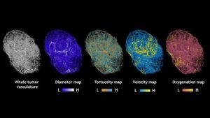
Map of tumors grown in mice
Using computation, researchers at Johns Hopkins are able to integrate images from in vivo MRI, ex vivo micro CT, and magnetic resonance microscopy to show the microvascular structure of human breast cancer tumors transplanted to mice. They describe the picture as being like a Google Maps of the tissue. This piece links to the full research article. MORE
Image Credit: Arvind Pathak, Ph.D., Johns Hopkins and ScienceDaily.com
