
Brain Mapping Called “Magnified Analysis of Proteome”
Magnified Analysis of Proteome (MAP)
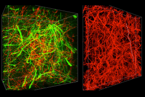
Neuron density in the human brain has made it nearly impossible to see the connections for impulse transmission with any kind of microscopy. To address that, biochemists at MIT have developed a technique they call “magnified analysis of proteome,” (MAP) which they describe here: “ The brain, being extremely dense, does not lend itself to optical microscopy techniques since the wavelength of light is too long to resolve many of the organ’s details. To overcome this, the researchers took an unexpected approach of simply stretching a preserved brain while keeping most of its characteristics intact. This is done by injecting acrylamide polymers inside the brain, turning it into a transparent gel-like object with all the proteins and neuronal connectivity intact. Along this process, the proteins are denatured and fluorescent antibody tags that stick to specific molecules are introduced.” MORE
Image Credit: MIT and MedGadget.com
