
Guiding Epidural Needles
August 9, 2016 | Terry Sharrer
Epidural Placement Guide
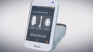
The epidural space of the spine exists between the vertebrae and dura, beneath which is the arachnoid space filled with cerebrospinal fluid under pressure. Thus it takes a skilled, steady hand to thread the needle through the ligaments without puncturing the fibrous dura. Scoliosis and calcification can complicate the needle placement; so, to improve this procedure, two biomedical engineering students at the University of Virginia have invented a hand-held devices that images the spine by ultrasound, and locates the best position for needle placement. After the needle is inserted, a catheter is put through the needle to deliver a pain-numbing drug. See the video. MORE WITH VIDEO
Image Credit: University of Virginia and Newsplex
