
Seeing Neurons as They Work
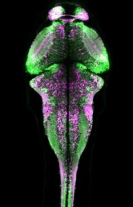
CaMPARI Fluorescence in a Larval Zebrafish
Calcium-sensitive fluorescent molecules have allowed researchers to see neuronal activity for some time, but mainly in a flash. Now, scientists at the Howard Hughes Medical Institute’s Janelia Research Park (Ashburn, VA) have found a way to display neuronal activity for a longer time in freely moving animals. As this piece describes it: “To make CaMPARI, the team started with a fluorescent protein called Eos. Eos emits a green fluorescence until it is exposed to violet light, which permanently alters the protein so that it fluoresces in red. That conversion from green to red gives us a permanent signal. So we just needed a way to couple that conversion to the activity that’s going on in the cell.” To do that, the scientists incorporated a calcium-sensitive protein known as calmodulin, which makes the color change dependent on the burst of calcium that accompanies neural activity. It’s the same domain that scientists added to fluorescent proteins to make calcium-responsive GCaMP sensors.” MORE
Image Credit: Looger Lab, HHMI/Janelia and MedicalNewsToday.com
