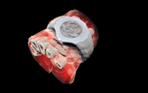
Compact Scanner Produces 3D X-Ray Images in Color
February 23, 2021 | Terry Sharrer

3D image of a wrist with a watch
With a detector chip from the European Organization for Nuclear Research (CERN) in Switzerland, a New Zealand company has developed a small x-ray system for scanning wrist and ankle joints. “Data collected by the scanner is processed with specialized algorithms to generate a 3D model. Specific densities are assigned different colors, so that bones appear white, muscle appears red and implants can be blue or green. During the feasibility trial, the images revealed failure of fractured scaphoid bones to heal and provided evidence of complications such as sclerosis, displacement and small bony fragments.” MORE
Image Credit: MARS Bioimaging Ltd


