
Microscopy Using UV Light for Diagnosis
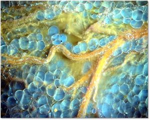
MUSE microscopy breast tissue
MUSE microscopy stands for Microscopy with Ultraviolet Surface Excitation, and is an imaging system from the University of California at Davis. It works in this way: “UV light at 280 nanometer spectral range illuminates about one square millimeter of specimen; Surface is limited to a few nanometers deep to make high-contrast images possible; Excitation light, at sub-300 nanometer spectral region, elicits bright emission from tissue specimens; Specimens, which were stained with conventional florescent dyes, emit photons; Photons are captured using glass-based microscope optics; A Python programing language solution, with a graphics unit, converts MUSE images in real-time; Images are comparable to the hematoxylin and eosin versions histologists and pathologists are accustomed to.” MORE
Image Credit: MUSE Microscopy Inc.
